


")
")
")


Research work carried out in this area essentially consists of applications of different radiations, in the most varied fields, among which we highlight:
- Non-Destructive Testing of Materials and Equipment;
- Nuclear Medicine and Radiology;
- Environment.
At the same time, other research works are developed, such as:
- Mathematical Data Analysis Models;
- Image processing;
- Nuclear Instrumentation;
- Development of Nuclear Detectors;
- Radiological Protection and Dosimetry.
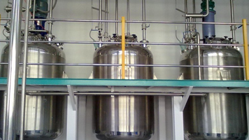
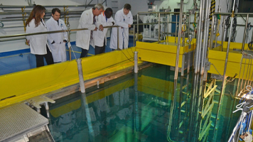
Research in the area of Applied Nuclear Physics is usually linked to other institutions in the nuclear area (e.g., Institute of Nuclear Engineering, Institute of Radioprotection and Dosimetry, Brazilian Center for Physics Research, Institute of Physics at UFRJ, Embrapa-CTAA, National Laboratory of Sincroton Radiation, etc.), but concentrates in the facilities of the Nuclear Instrumentation Laboratory (LIN / COPPE).
The area has been developing techniques for detecting and applying nuclear radiation for several years. Many research works result from master’s and doctoral theses dealing with radiography, image reconstruction, special detectors, etc., accumulating their own technology. Recent advances and multiple applications of computed tomography, together with several institutions and researches that have sought the LIN / COPPE, led us to launch a computerized tomography program, qualified to develop:
- All image reconstruction “software” technology;
- An understanding of the “hardware” technology needed for tomographic systems;
- Prototypes of computerized tomographs, with a high level of national technology, versatile enough to, through small changes, be competent in several areas.
The major results of the project will be both the industrialization, by national companies, of high-tech equipment, but at a much lower cost than imported ones, as well as an elimination of chaos (technical and financial) resulting from its maintenance by multinational companies. In addition, the greatest triumphs will be the training of personnel in state-of-the-art technology and the interaction of research groups in related areas with the national industry, the final objective of the project: the prototype of an integral tomographic system.
The global project of the tomographic system is broken down into several subprojects, according to the following lines of research:
Neutron radiography, gamma radiation and X-rays
- Determination of radiographic parameters of various detection systems (converters and films) used to obtain neutron radiographies (neutrongraphies);
- Definition of experimental conditions for neutrongraphy of objects that require quality Non-Destructive Testing;
- Detection of faults by neutrongraphy in ceramic fine occurrences;
- Assessment of the detection limit of defects in materials;
- Sensitivity of neutrongraphy in detecting corrosion products in aircraft;
- Study of the feasibility of neutrongraphy in biological tissues of the human and/or animal body;
- Determination of real-time radiography system operating parameters;
- Real-time scanning of radiographic images;
- Development of tomographic systems based on radiographies;
- Assembly of a transportable neutrongraphic system;
- Morphological characterization and quantitative analysis of bacteria in vitro;
- Detection of drugs and explosives.
Computed tomography with radiation
- Study of the fundamentals of reconstruction used in three-dimensional analysis of object structure through measurements of angular projections, using X-rays, gamma, or neutrons;
- Use of these methods for assembling experimental systems, which serve as a prototype of a computerized tomograph for use in industrial tests and medical diagnostics.
Compton scattering
- Research on the fundamental mechanism of gamma radiation scattering in matter (Compton effect) and application of this phenomenon in industry, in Non-Destructive Testing of materials and in medicine, in the reconstruction of images of various organs of the human body;
- Assembly of a prototype to serve as a model for fully automated apparatus.
Diffraction
- Study of radiation diffraction parameters in crystalline and amorphous materials for application in diffraction tomography, with use in industrial and medical areas.
Detection Systems
- Study and development of detectors based on scintillator crystal-photodiode coupling for construction of linear arrays applied in inspection systems;
- Development of fast, charge-sensitive preamps for use in photodiode systems.
Research involving Electronic Paramagnetic Resonance (EPR)
- Study and development of techniques for identifying irradiated foods, especially fruits and products with high moisture content, which makes the identification process complex;
- Research of new dosimeters for applications in industrial dose irradiation, mainly in sterilization irradiators or for food irradiation;
- Research of new dosimeters for the evaluation of absorbed doses in nuclear accident, using (RPE), through the evaluation of induced radicals in several ceramic materials and bioapatites.
Quantitative Measurements by X-Ray Fluorescence
- Application of the energy dispersive X-ray fluorescence technique in the measurement of trace elements in air fresheners for measurement of source and contamination at the ppm and ppb level.


